


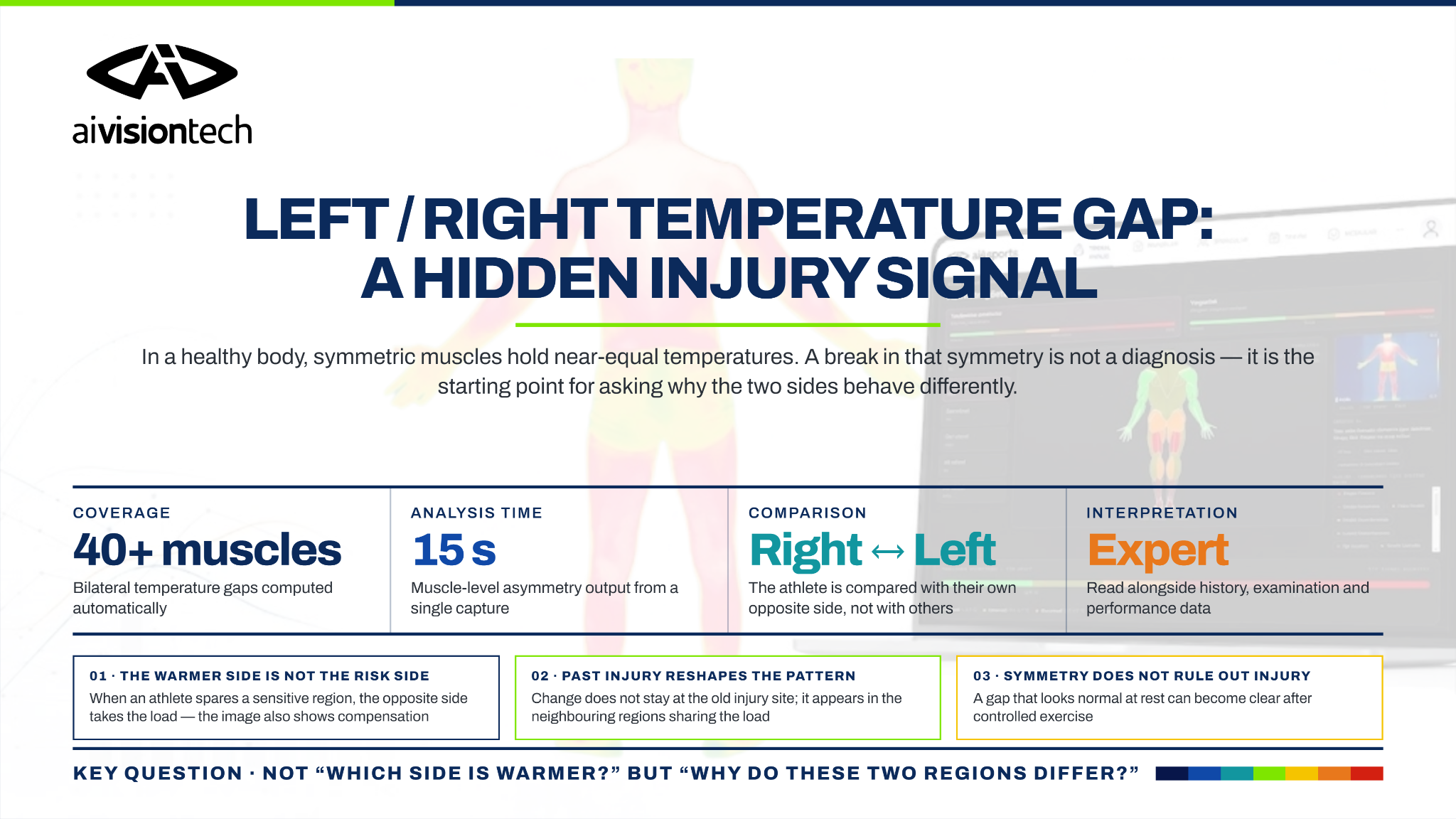
In a healthy body, homeostatic mechanisms generally produce similar thermal radiation across corresponding anatomical regions. Under normal conditions, skin temperatures on opposite sides of the body are therefore expected to be equal or very close. Thermal asymmetry analysis can make unexpected differences between an individual's right and left muscle groups visible. A large temperature difference, however, must not be used as a standalone diagnosis of injury. Dominance, training load, previous injury, circulation, and autonomic nervous system activity can all influence the same thermal image. Thermal asymmetries should therefore be interpreted by an expert in the context of the athlete's history and anatomy.
With a single ai4sports thermal analysis, asymmetry values across 40+ muscle groups can be assessed in less than 15 seconds. In thermal asymmetry, the region at risk should not automatically be identified as the side with the higher temperature; the warmer muscle region is not always the primary problem area.
Why Do the Right and Left Sides Usually Have Similar Temperatures?
The human body is not perfectly geometrically symmetrical. Nevertheless, corresponding anatomical regions on the right and left are influenced by similar circulatory, metabolic, and thermoregulatory mechanisms. For this reason, healthy conditions are generally associated with similar skin temperatures on both sides of the same muscle group.
This characteristic is a major advantage in thermography. Instead of comparing one athlete's right hamstring with another athlete's hamstring, the athlete's own right and left hamstring regions can be compared with each other. This reduces the influence of variables such as age, body composition, overall metabolism, and individual thermal profile.
Uematsu and colleagues examined corresponding anatomical regions in healthy individuals and showed that bilateral temperature differences were generally small [1]. A normative study by Niu and colleagues in another population also supported the tendency toward right-left thermal symmetry in healthy individuals [2].
For this reason, the core measurement logic in a single ai4sports thermal analysis is to evaluate the temperature difference between the right and left sides of the same anatomical muscle region.
What Does Thermal Asymmetry Show?
Thermal asymmetry is a signal that two symmetrical anatomical regions are not displaying the same physiological behavior.
There is no single explanation for such a difference. Changes in local blood flow, inflammatory processes, autonomic nervous system responses, dominant-side use, previous injuries, and rehabilitation can all alter skin-temperature distribution.
A thermographic difference should therefore not be interpreted directly as “This muscle is injured.” A more appropriate question is “Why are these two symmetrical regions behaving differently?”
Recent systematic reviews of thermography in football report thermal asymmetry as one of the variables used when evaluating injury risk, fatigue, and physiological load. At the same time, these studies emphasize that measurement conditions and clinical context must be considered during interpretation [3].
Thermography therefore does not show anatomical damage itself; it shows the physiological response reflected at the body surface.
Is the Warmer Side Always the Risk Side?
No.
One of the most common errors in thermal analysis is to assume that the warmer region in an asymmetry is automatically the problematic region.
Higher temperature may be associated with increased local blood flow, an inflammatory response, or greater muscle activity. Yet the clinically meaningful region is not always the warmer one.
In a study by Uematsu and colleagues involving patients with low-back pain and sciatica, the affected lower extremity was significantly cooler than the opposite side [4]. This study was not conducted on sports injuries, but it is important for understanding basic thermal physiology: thermal disturbance does not present only as an increase in temperature.
Load distribution can also change in athletes. An athlete may unconsciously use a sensitive or previously injured region less. In that case, muscle groups on the opposite side can take on more load.
As a result, the warmer side on a thermal image may not always be the primary problem area. The image may also be showing the result of compensation.
How Can a Previous Injury Change Thermal Distribution?
Previous injuries can alter an athlete's movement pattern. Even after pain has fully resolved, muscle use, joint movement, or load distribution may remain different from the pre-injury state.
A study by Bayrak, Çevik, and Ceylan compared U19 footballers with a history of ankle injury with players without an injury history. The study evaluated not only the ankle region but also the patellar tendon, tibialis anterior, and medial-lateral calf regions [5].
In players with an injury history, thermal changes were not limited to the original injury site. Changes in asymmetry were also observed in other lower-extremity regions.
After an ankle problem, an athlete may redistribute load differently. Thermal changes may therefore appear not only at the ankle but also in patellar or calf regions that share the load.
In a single thermal analysis, the focus should therefore not be limited to the hottest or apparently highest-risk point; the distribution across the entire kinetic chain should also be considered.
Can Dominant-Side Use Create Asymmetry?
Yes. In sports that rely heavily on unilateral techniques, the dominant side can naturally be loaded differently.
Archery is a clear example. In a study of 30 archers by Sanchis-Sanchis and colleagues, thermal asymmetry increased in the upper back, posterior shoulder, arm, and elbow regions after a simulated competition. In some regions, the bow side showed a higher temperature [6].
Another study in padel players reported post-training temperature increases in the forearm, shoulder, and arm of the dominant side [7].
These findings show that unilateral loading can be reflected in thermal distribution. However, it would be incorrect to conclude that the dominant side will always be warmer.
A study in cyclists found mechanical differences in right-left crank torque without a corresponding degree of thermal asymmetry [8]. Artificially induced mechanical asymmetry in runners likewise did not produce a significant thermal asymmetry in plantar temperature in the same direction [9].
The key distinction is therefore that mechanical asymmetry and thermal asymmetry are not the same thing.
A thermal image reflects not only how much a muscle works, but also the physiological consequence of that loading on circulation and thermoregulation.
Studies reporting thermal asymmetry associated with dominance generally evaluate images obtained after competition or training. It should also be remembered that normal thermoregulation is expected to recover after an appropriate recovery period.
Why Can the Same Injury Look Thermally Different in Different Athletes?
Because athletes do not all produce the same physiological response to the same load.
Bayrak, Ergene, and Ceylan studied two elite footballers who returned to sport after proximal rectus femoris injury. The athletes were exposed to similar training loads, yet their thermal responses differed [10].
The athlete who returned after surgical treatment showed a greater change in right-left temperature difference and sustained a reinjury in the same region during the following two weeks.
Because this report included only two cases, it cannot be interpreted as proof that thermography predicted reinjury. It does, however, demonstrate an important point: the same diagnosis, the same training load, and a similar return-to-sport period do not necessarily produce the same thermal response.
Knowledge of the athlete's injury history is therefore an important part of interpreting a single thermal analysis.
Does Thermal Symmetry Mean There Is No Injury?
This question cannot be answered with a simple yes or no. Thermography should be understood as an adjunctive risk indicator, not a diagnostic test. Thermal imaging measures heat emitted from the surface of the skin, while physiological processes in deep muscle groups or ligamentous regions may not be directly visible at the skin surface.
A similar appearance on the right and left sides does not prove that the athlete is injury-free.
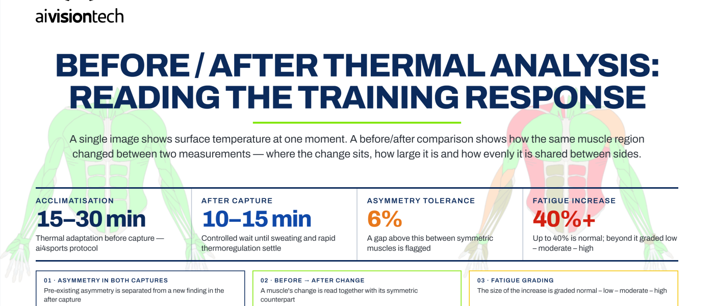
A sartorius case report by Bayrak, Ergene, and Ceylan provides a useful example. Initial resting thermal imaging of a professional footballer showed no clear asymmetry. After controlled exercise, however, a thermal difference in the sartorius region became apparent. The anatomical injury was subsequently confirmed by MRI [11].
This case clearly illustrates a limitation of thermography. Normal thermal symmetry is not a test that rules out injury. Likewise, high thermal asymmetry does not confirm an injury by itself.
Between these two extremes, thermography provides decision-support data that can identify regions requiring closer assessment.
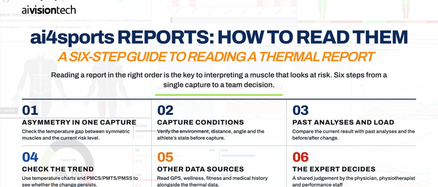
How Should a Single Right-Left Difference Be Read?
When a single thermal image is analyzed with ai4sports, bilateral temperature differences across 40+ muscles and anatomical regions are calculated automatically.
The report, however, should not be interpreted only through its risk colors. The following questions should be considered first:
- Which anatomical region shows a right-left difference?
- What is the direction of the temperature difference?
- Has the athlete previously been injured in this region?
- Which side is dominant?
- Does the sport naturally create asymmetrical loading?
- Is the athlete in rehabilitation or a return-to-play process?
- Do neighboring muscle groups show a similar distribution?
- Is there pain, tenderness, or restricted movement?
It is also important not to equate thermal asymmetry with strength asymmetry. In a study of healthy football players, thermal asymmetries were present in a substantial proportion of athletes, yet those differences were not directly associated with muscular strength imbalances [12].
Thermal analysis therefore does not prove a specific mechanical or anatomical cause on its own. Its main value is showing which muscle region warrants investigation and asking why it is behaving differently.
Conclusion: Thermal Asymmetry Is a Starting Point, Not a Final Answer
Similar temperatures in right and left muscle groups are a fundamental feature of human thermoregulation. A disruption in that symmetry indicates that the two sides are not behaving in the same physiological way.
There is no single reason for that difference. A warmer side may reflect local loading or an inflammatory response. A cooler side may be associated with circulatory or autonomic changes. Heavy use of the dominant side can create a natural difference. A previous injury may also cause the athlete to shift load to the opposite side or to neighboring muscle groups.
For this reason, the goal of a single ai4sports thermal analysis is not to answer “Which side is warmer?” The more important question is “Why are these two symmetrical muscle regions behaving differently?”
The thermal image shows where that investigation should begin. The answer should be completed by a healthcare professional using the athlete's history, physical examination, performance data, and, when needed, other imaging methods such as MRI or ultrasound.
References
- Uematsu, S., Edwin, D. H., Jankel, W. R., Kozikowski, J., & Trattner, M. (1988). Quantification of thermal asymmetry. Part 1: Normal values and reproducibility. Journal of Neurosurgery, 69(4), 552–555. https://doi.org/10.3171/jns.1988.69.4.0552
- Niu, H. H., Lui, P. W., Hu, J. S., Ting, C. K., Yin, Y. C., Lo, Y. L., Liu, L., & Lee, T. Y. (2001). Thermal symmetry of skin temperature: Normative data of normal subjects in Taiwan. Zhonghua Yi Xue Za Zhi (Taipei), 64(8), 459–468.
- Barajas Ramón, Y., Calleja-González, J., Luaces-Carreño, J., & Velarde-Sotres, Á. (2026). Infrared thermography to assess fatigue, injury risk factors and recovery in soccer: A systematic review of original studies. Frontiers in Physiology, 17. https://doi.org/10.3389/fphys.2026.1835464
- Uematsu, S., Jankel, W. R., Edwin, D. H., Kim, W., Kozikowski, J., Rosenbaum, A., & Long, D. M. (1988). Quantification of thermal asymmetry. Part 2: Application in low-back pain and sciatica. Journal of Neurosurgery, 69(4), 556–561. https://doi.org/10.3171/jns.1988.69.4.0556
- Bayrak, A., Çevik, M., & Ceylan, M. (2026). Thermal asymmetry in football players following ankle injury: Findings related to training load. In Artificial Intelligence over Infrared Images for Medical Applications (AIIIMA 2025), Lecture Notes in Computer Science, Vol. 16308, 128–142. Springer. https://doi.org/10.1007/978-3-032-10990-3_9
- Sanchis-Sanchis, R., Priego-Quesada, J. I., Ribas-Garcia, V., Carpes, F. P., Encarnacion-Martinez, A., & Perez-Soriano, P. (2020). Effects of asymmetrical exercise demands on the symmetry of skin temperature in archers. Physiological Measurement, 41(11), 114002. https://doi.org/10.1088/1361-6579/abc020
- Gómez-Carmona, P. M., et al. (2024). Preliminary Application of Infrared Thermography to Monitoring of Skin Temperature Asymmetries in Professional Padel Players. Sensors, 24(14), 4534. https://doi.org/10.3390/s24144534
- Trecroci, A., Formenti, D., Ludwig, N., Gargano, M., Bosio, A., Rampinini, E., & Alberti, G. (2018). Bilateral asymmetry of skin temperature is not related to bilateral asymmetry of crank torque during an incremental cycling exercise to exhaustion. PeerJ, 6, e4438. https://doi.org/10.7717/peerj.4438
- Gil-Calvo, M., Herrero-Marco, J., González-Peña, R. J., Perez-Soriano, P., & Priego-Quesada, J. I. (2020). Acute effect of induced asymmetrical running technique on foot skin temperature. Journal of Thermal Biology, 91, 102613. https://doi.org/10.1016/j.jtherbio.2020.102613
- Bayrak, A., Ergene, M. C., & Ceylan, M. (2023). Monitoring the Reactions of Athletes With History of Rectus Femoris Proximal Tear Healed With Different Methods to Training Load With Thermography. Turkish Journal of Sport and Exercise, 25(2), 231–239. https://doi.org/10.15314/tsed.1292549
- Bayrak, A., Ergene, M. C., & Ceylan, M. (2024). Thermography method under the influence of exercise in the detection of muscle injuries: Sartorius muscle case report. Journal of Bodywork and Movement Therapies, 39, 109–115. https://doi.org/10.1016/j.jbmt.2024.02.029
- Teixeira, R. M., et al. (2020). Muscular Strength Imbalances Are Not Associated with Skin Temperature Asymmetries in Soccer Players. Life, 10(7), 102. https://doi.org/10.3390/life10070102