


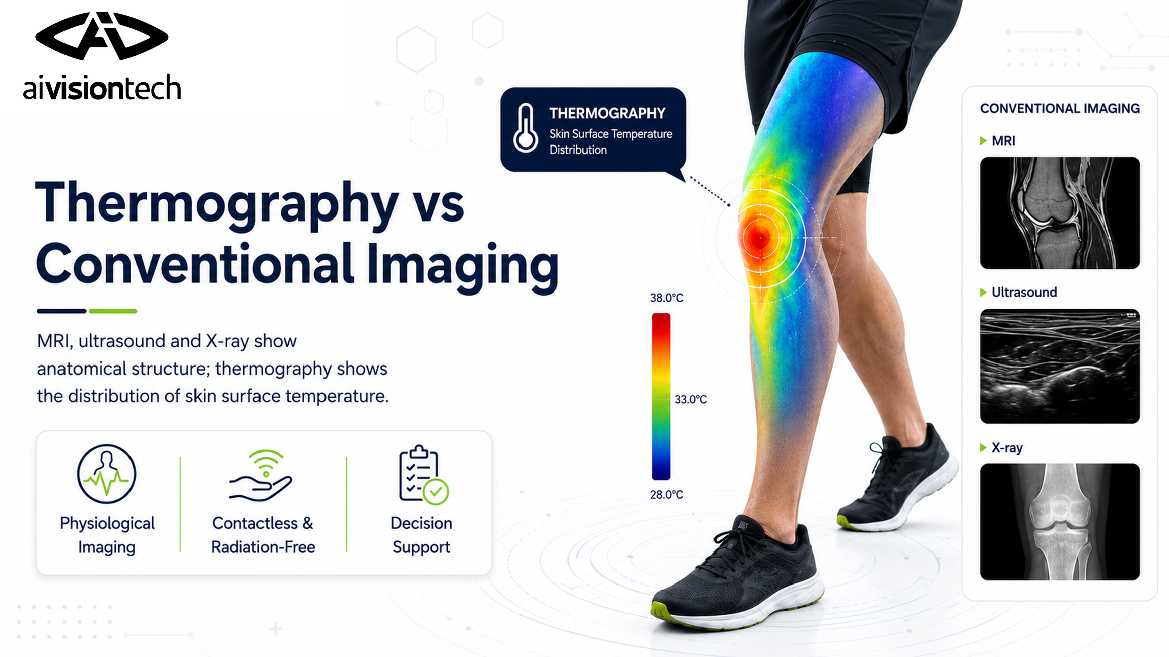
Thermography evaluates infrared energy emitted by the body, while conventional imaging methods evaluate anatomical structure.
While imaging systems such as MRI, ultrasound and X-ray provide information about tissue structure, thermography examines temperature distribution by measuring infrared energy at the skin surface. For more detailed information about thermography, see What Is Thermal Imaging and How Is It Used in Sports?. Thermography is therefore not an alternative system intended to replace conventional imaging methods; instead, especially because repeated imaging can be performed, it provides a different data layer that can complement them in periodic monitoring and the assessment of physiological change, supporting decision-making.
What Do Imaging Methods Measure?
When comparing medical imaging methods, the first question should not be “which device is better?” but “what does each device measure?” Each imaging system is used for different purposes.
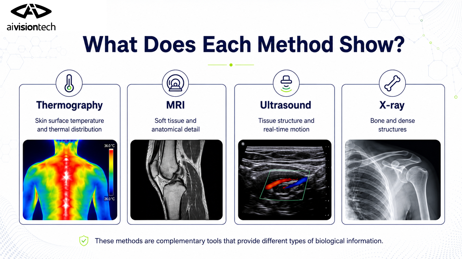
X-ray is based on the fact that X-rays pass through tissues in different amounts. Dense structures such as bone absorb more radiation, making X-ray particularly useful for visualizing fractures, cracks and certain joint abnormalities. MRI uses a strong magnetic field and radiofrequency signals to display the anatomical structure of muscles, tendons, ligaments, cartilage and other soft tissues in high detail. Ultrasound uses the reflection of ultrasonic sound waves from tissues. It can be used to assess the structure of muscles, tendons and some superficial tissues dynamically and in real time.
Thermography does not directly image human anatomy. See What Is Thermal Imaging and How Is It Used in Sports?. A thermal camera detects infrared radiation emitted by human skin and converts it into temperature values. It assigns a temperature value to each pixel and produces a temperature map. This map provides information about the physiological response reflected at the skin surface. The important point is that thermography does not directly show a deep muscle tear, ligament rupture or bone lesion. For this reason, an asymmetry seen in a thermal image is not a structural diagnosis; it is a physiological finding used in the assessment of injury-related and inflammatory responses.
For thermal imaging to be used in sport and exercise settings, the imaging environment, participant preparation, camera position and analyzed anatomical regions need to be standardized. The international TISEM approach also emphasizes that measurement conditions should be controlled and repeatable to allow reliable comparison of skin temperature [1]. The ai4sports, ai4body, ai2neo and ai4animal products developed by aivisiontech also provide users with appropriate imaging environment protocols based on this guidance for suitable analyses.
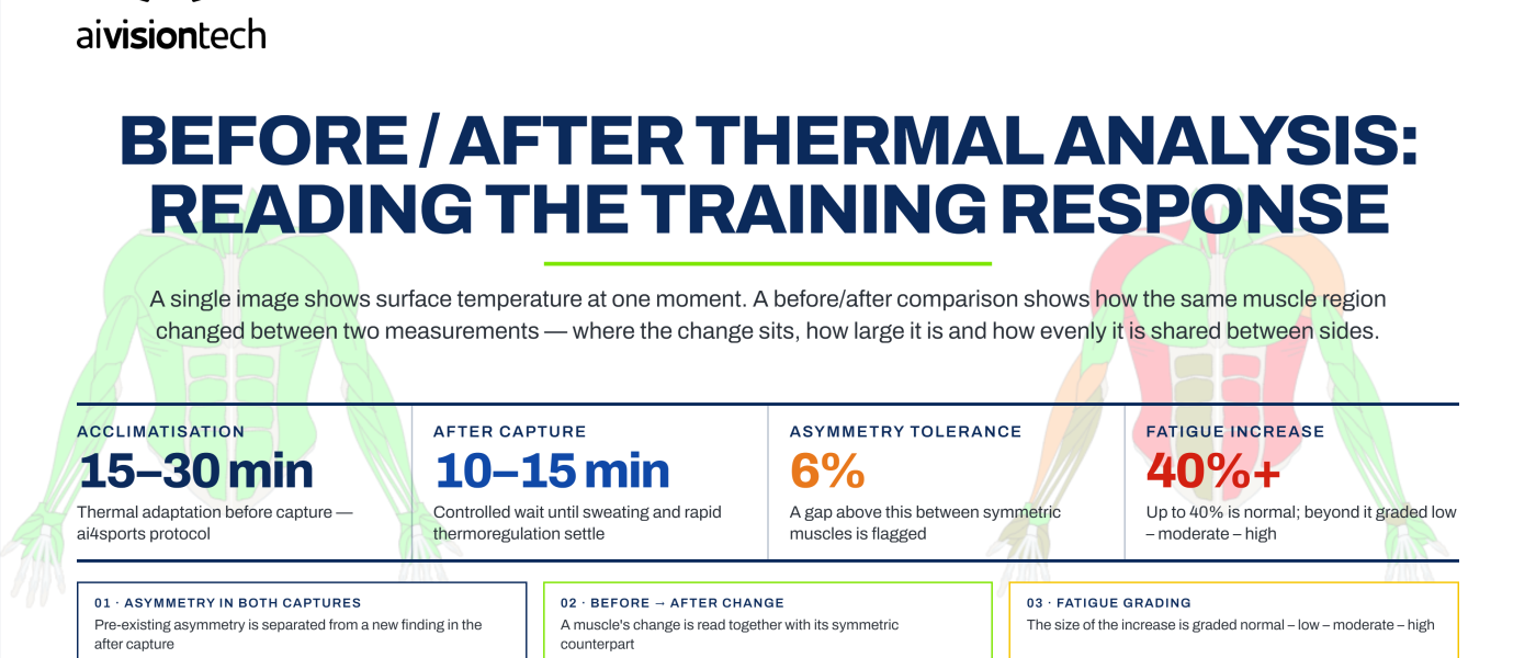
ai4sports carries this distinction in thermal imaging directly into its analysis logic. Instead of interpreting a thermal image as though it were an MRI-like anatomical image, it evaluates temperature values before and after training, differences between symmetrical muscle groups and the individual’s own historical thermal measurements. The result is not presented as a diagnosis, but as a structured decision-support data point and risk indicator for coaches, physiotherapists and healthcare teams.
Comparison of Thermography, MRI, Ultrasound and X-ray
When the same anatomical region is examined with different imaging methods, different questions are actually being answered. In the knee, for example, X-ray focuses on bone and joint structures; MRI on the meniscus, ligaments, cartilage and other soft tissues; ultrasound on superficial tendons and muscle structures; and thermography on the distribution of high and low temperatures at the skin surface and right-left thermal differences.
| Feature | Thermography | MRI | Ultrasound | X-ray |
|---|---|---|---|---|
| What does it show? | Skin surface temperature and thermal distribution | Anatomical structure and soft tissue | Tissue structure and movement | Primarily bone and dense structures |
| Ionizing radiation | None | None | None | Yes |
| Dynamic / real-time use | Yes, for surface temperature | Limited in routine use | Yes | No in standard radiography |
| Suitability for frequent repeated measurement | Very high | Limited by time and cost | High | Not suitable because of radiation exposure |
| Main focus | “Is there a thermal change in the skin?” | “Is there a structural change in the tissue?” | “What does the superficial structure look like?” | “Is there an abnormality in bone structure?” |
| Limitations | Does not directly show deep tissue | Longer acquisition, cost and access | Operator- and acoustic-window-dependent | Limited soft-tissue detail |
Examples in the literature also show that these methods can complement one another in the same clinical question. In knee osteoarthritis, an association has been reported between differences in the patellar region temperature map and radiographic disease severity, demonstrating that thermography and X-ray may show different aspects of the same clinical condition even though they do not measure the same thing [2]. Similarly, in the patellar tendon, mechanical properties assessed with ultrasound elastography and surface temperature assessed with thermography were examined together within the same evaluation, and a positive overlap was observed [3].
Therefore, the appropriate question is not “thermography or MRI?” but “what biological information is needed in this person’s evaluation?” When structural damage is suspected, MRI, ultrasound or X-ray may be required; thermography may provide an additional layer for monitoring physiological response and load-related changes during the same process.
Learn how thermal images are acquired in What Is Thermal Imaging and How Is It Used in Sports?.
What Are the Advantages of Thermography?
The most evident advantage of thermography is that it provides a contactless and radiation-free (non-invasive) measurement. The device does not transmit radiation energy to the person; it measures infrared energy naturally emitted from the skin surface. Therefore, when used with an appropriate protocol, the same person can be imaged repeatedly.
This is particularly important for athlete monitoring during a season, return-to-play and rehabilitation processes. A single MRI or ultrasound assessment shows structural status at a specific point in time, whereas thermal analysis can be repeated on different days or before and after training to monitor the direction of temperature change. The demonstration that thermal asymmetry in the knee region changes over time during ACL rehabilitation in professional and semi-professional football players supports the suitability of thermography for serial measurements during rehabilitation [4].
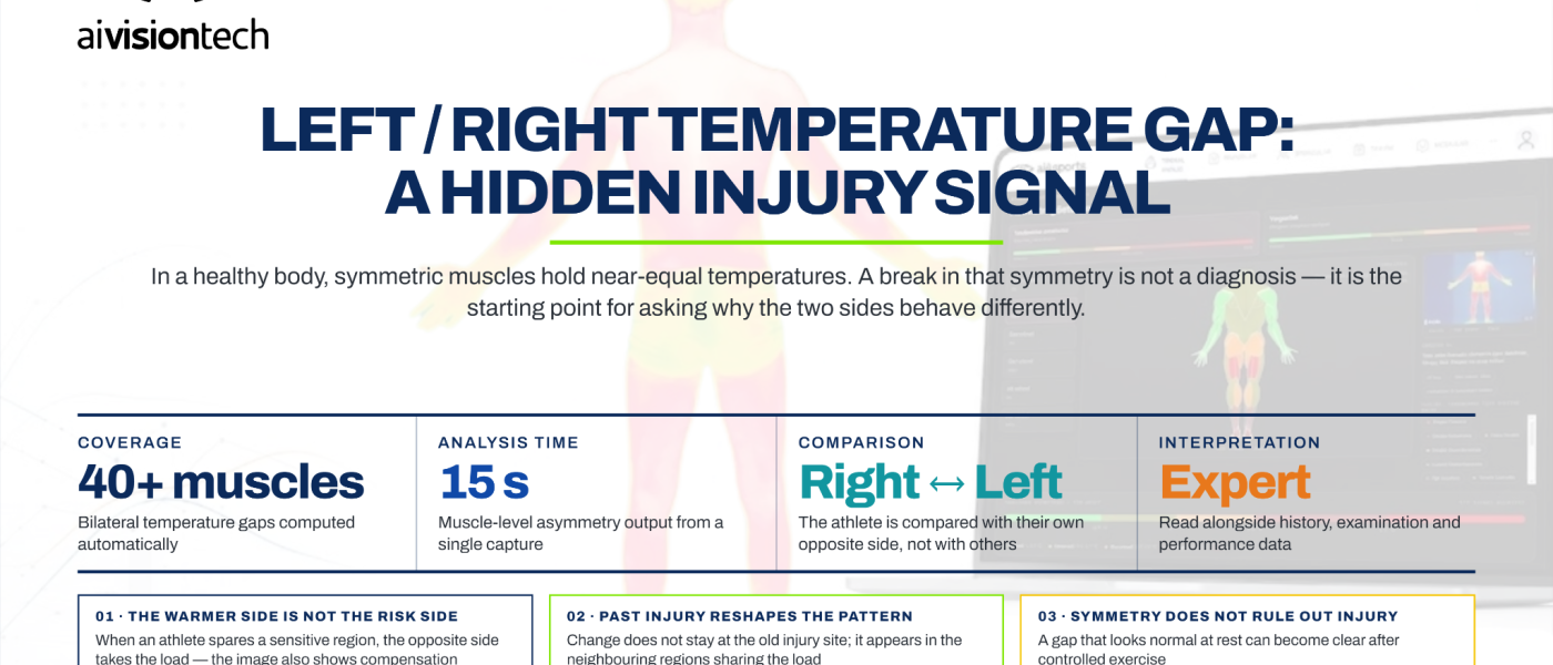
In ai4sports, more than 40 muscle groups are automatically segmented from the thermal image, allowing a transition from image acquisition to structured analysis in under 15 seconds. To date, more than 2 million thermal muscle-group analyses have been processed on the system. This scale is the practical counterpart of using thermography not as a single clinical image, but as repeated, multi-region monitoring data.
The same approach is brought to the sideline with ai4sportsGO. Imaging can be performed with a mobile thermal camera connected to a phone via USB Type-C; regional findings and scores can be reviewed on the mobile device through the ai4sportsGO application. This allows thermal analysis to be used not only in a fixed imaging area but also, when standardized imaging conditions are maintained, in field or performance-center settings.
What Are the Limitations of Thermography?
Knowing what thermography cannot do is as important as knowing what it can do. The main limitation is that a thermal camera does not directly image deep tissue. The measured value is skin surface temperature. Therefore, the grade of a muscle tear, a meniscus injury, ligament rupture or a bone lesion cannot be structurally diagnosed from a thermal image.
Thermography should be positioned as a risk-assessment and decision-support tool, not as a diagnostic tool.
A second important point is that skin temperature is not affected only by injury. Room temperature, humidity, air flow, sunlight, exercise, sweating, clothing, creams, recent showering and insufficient acclimatization to the environment can all change the measurement. A thermographic examination is therefore not simply an arbitrary photograph; it requires a standardized protocol [1].
Thermal asymmetry does not by itself indicate pathology either. Although prospective and follow-up studies in athletes using thermography have shown useful associations between temperature asymmetries and injury or rehabilitation processes, thermography alone does not indicate which tissue is damaged or the anatomical severity of the damage [4],[5]. Thermal asymmetries should be treated as a risk indicator, while the underlying cause and clinical diagnostic process should be evaluated using gold-standard imaging methods.
For this reason, the core approach in aivisiontech products is clear: thermal analysis provides decision support; it does not replace professional medical diagnosis. The clinical significance of a finding should be evaluated by a healthcare professional together with symptoms, physical examination, athlete history, performance data and, when necessary, MRI, ultrasound or X-ray imaging.
How Is a Thermographic Examination Performed?
The first step toward reliability in thermographic examination begins before the image is taken. The person should be kept away from sudden temperature changes and allowed to adapt to the imaging environment. Room temperature and humidity should be kept as stable as possible, and direct sunlight or strong air flow should be avoided. TISEM recommendations used in sport and exercise assessments specifically emphasize standardization of participant preparation, environmental conditions, camera settings, image acquisition and data analysis [1].
During imaging, the distance between camera and participant, viewing angle and body position should be kept as consistent as possible across measurements. An appropriate emissivity value for human skin (0.98) should be used, and the anatomical regions being compared should be imaged in the same position. Particularly when right-left temperature differences are being evaluated, capturing both sides in the same frame or with the same standardized imaging protocol improves comparability.
At the evaluation stage, it is not correct to interpret only the colors visible in the image. The same color may represent different temperature values under different minimum-maximum temperature scales. Interpretation should therefore be based on the region’s numerical mean temperature, maximum/minimum values, bilateral differences and change compared with previous measurements.
In the ai4sportsGO workflow, the process begins with acquisition of a thermal image using a mobile thermal camera attached to the phone. After image acquisition, regional analyses and scores can be reviewed on the mobile device. This accelerates the transition from measurement to report while preserving a standardized imaging protocol suitable for field use. The mobile application is also synchronized with ai4sportsHUB, allowing users to review analyses performed in ai4sportsGO in the web environment as well.
How Are These Methods Used Together in Sports Medicine?
The strongest approach in sports medicine is to use imaging methods not as competitors, but as tools that answer different questions. For example, during an intensive training period, thermography can be used for rapid, broad screening across a squad. If an unusual thermal change between right-left muscle groups or a marked deviation from the athlete’s own baseline is observed, the healthcare team can assess the athlete through history and physical examination. If a structural problem is suspected, the process can continue with ultrasound, MRI or, when appropriate, X-ray.
This complementary approach is also supported in the literature. In a patellar tendon study, the mechanical properties measured by ultrasound elastography and the temperature measured by thermography were treated as different biological variables within the same protocol [3]. In knee osteoarthritis, the relationship between thermal measurements and radiographic findings was examined, showing that surface temperature and structural imaging can provide different yet potentially related information [2].
ai4sportsHUB is the layer that facilitates information sharing within the team. Squad overview, risk body map and periodic scores such as PMCS, PMSS and PMTS can be followed within the same structure by coaches, physiotherapists and healthcare staff. ai4sportsGPT can generate decision support using the athlete’s analysis history and current findings. Regardless of the imaging method, however, clinical decisions and diagnosis remain the responsibility of healthcare professionals.
For this reason, the most appropriate role of thermography in sports medicine is not to be the first and only diagnostic method, but to complement existing clinical workflows through rapid screening, periodic follow-up and structured monitoring of physiological change.
Frequently Asked Questions (FAQ)
Can thermography replace MRI?
No. MRI and thermography measure different biological information. MRI visualizes the anatomical structure of muscles, tendons, ligaments, cartilage and other soft tissues in detail, while thermography measures temperature distribution at the skin surface. A thermal asymmetry is not the direct equivalent of structural damage that may be visible on MRI. Thermography can be used as a complementary tool to support referral for MRI when appropriate or to follow physiological change during rehabilitation.
What are the advantages of thermography?
It is contactless, does not involve ionizing radiation, can be applied quickly and can be repeated frequently in the same person. These features make it suitable particularly for right-left temperature comparison, pre/post-training monitoring and tracking thermal changes over time during rehabilitation.
How is a thermographic examination performed?
First, the person should be prepared for imaging in a controlled environment and allowed to adapt to the room temperature. Images are then acquired after standardizing parameters such as camera distance, angle, position and emissivity. Evaluation should not be based only on the color map, but on numerical temperature values, right-left differences and, where possible, comparison with the individual’s previous measurements [1].
In which hospitals is thermography available?
Thermography is not as routinely available in every healthcare institution as MRI or X-ray. Its use and device availability vary by institution; it may be found in sports medicine, physical medicine and rehabilitation, vascular assessment or certain research units. For a specific hospital, the most accurate information should be obtained from that institution’s imaging department or relevant clinical unit.
When does sports medicine become involved?
Sports medicine can be involved in the evaluation of acute injuries, recurrent pain and overuse problems, return-to-sport processes, training-load management and performance-related health problems. Thermography may be one of the assessment tools used by the healthcare team in these processes and, when necessary, is considered together with physical examination, laboratory tests and other imaging methods.
How reliable are thermography results?
The reliability of thermographic measurement depends on the camera used, imaging protocol, environmental conditions and analysis method. Under standardized conditions, skin temperature and bilateral temperature differences can be measured reproducibly; however, the clinical meaning of a thermal finding should not be derived from the image alone [1]. Therefore, a reliable temperature measurement and a reliable medical diagnosis are not the same thing.
Looking at the Same Body Through Different Windows
MRI, ultrasound, X-ray and thermography are not methods that give different answers to the same question; each makes a different property of the body visible. Conventional imaging methods reveal anatomical and structural changes, whereas thermography monitors physiological temperature distribution at the skin surface. The value of thermography therefore lies not in replacing the other methods, but in adding a data layer that they do not provide through rapid and repeatable measurements. The right question is not which imaging method is better, but which information is needed at which stage.
References
- Moreira, D. G., Costello, J. T., Brito, C. J., Adamczyk, J. G., Ammer, K., Bach, A. J. E., et al. (2017). Thermographic imaging in sports and exercise medicine: A Delphi study and consensus statement on the measurement of human skin temperature. Journal of Thermal Biology, 69, 155–162. https://pubmed.ncbi.nlm.nih.gov/29037377/
- Denoble, A. E., Hall, N., Pieper, C. F., & Kraus, V. B. (2010). Patellar skin surface temperature by thermography reflects knee osteoarthritis severity. Clinical Medicine Insights: Arthritis and Musculoskeletal Disorders, 3, 69–75. https://pubmed.ncbi.nlm.nih.gov/21151853/
- Cuevas-Cervera, M., et al. (2023). Patellar Tendon Elasticity and Temperature Following after a Single Session of Dry Needling in Individuals with Patellar Tendinopathy: A Randomized Controlled Trial. https://pubmed.ncbi.nlm.nih.gov/37761343/
- Bayrak, A., Ergene, M. C., & Ceylan, M. (2023). Monitoring the reactions of athletes with history of rectus femoris proximal tear healed with different methods to training load with thermography. Turkish Journal of Sport and Exercise, 25(2), 231–239. https://dergipark.org.tr/en/pub/tsed/article/1292549
- Bayrak, A., Ergene, M. C., & Ceylan, M. (2024). Thermography method under the influence of exercise in the detection of muscle injuries: Sartorius muscle case report. Journal of Bodywork and Movement Therapies, 39, 109–115. https://doi.org/10.1016/j.jbmt.2024.02.029